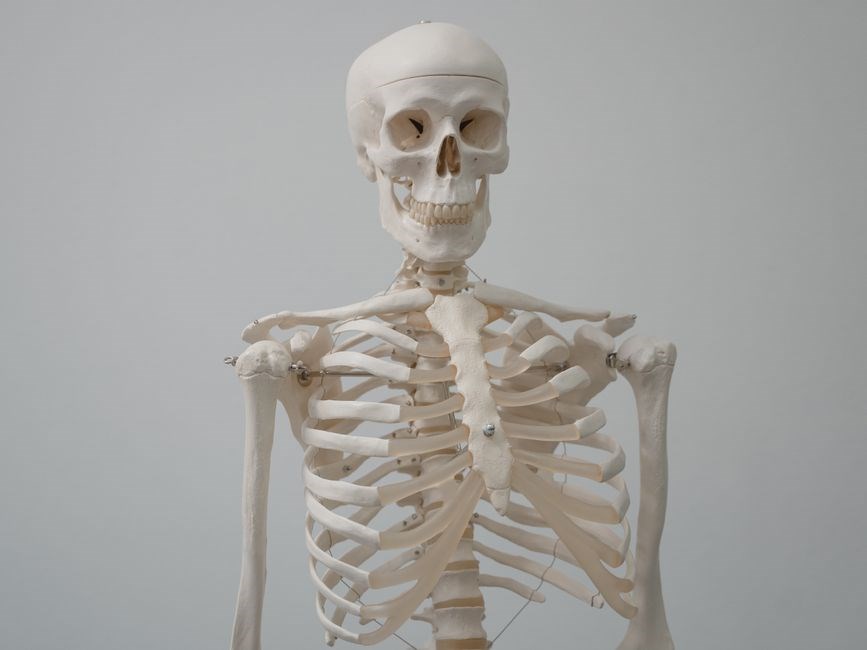
Overview of Musculoskeletal System
The musculoskeletal system integrates bones, joints, muscles, ligaments, tendons, and connective tissue, forming a dynamic framework that supports structure, protects organs, stores minerals, and facilitates movement through coordinated contraction and joint articulation.
Movement relies on it.
Definition and Components
The musculoskeletal system is the integrated network of bones, joints, muscles, ligaments, tendons, cartilage, fascia, and other connective tissues that together provide structural support, enable locomotion, protect vital organs, store minerals, and facilitate metabolic and endocrine functions. Its primary components include:
- Bone: A mineralized connective tissue composed of cortical (compact) and trabecular (spongy) bone, forming the rigid framework.
- Cartilage: A flexible, avascular tissue that cushions joints and shapes cartilage surfaces.
- Synovial fluid: A viscous liquid that lubricates synovial joints, reducing friction during movement.
- Ligaments: Fibrous bands that connect bone to bone, stabilizing joints.
- Tendons: Connective strands that attach muscle to bone, transmitting force for movement.
- Muscle fibers: Contractile cells grouped into skeletal muscle, responsible for voluntary motion.
- Fascia: Dense connective tissue that surrounds muscles, organs, and nerves, providing structural integrity.
- Nerves and blood vessels: Supply innervation and nutrients, enabling coordinated function.
Together, these elements form a dynamic system that balances rigidity and flexibility, allowing precise, efficient motion while maintaining structural integrity and protecting internal organs.
Clinical assessment of this system often employs imaging modalities such as X‑ray, MRI, and CT scans, alongside physical examination and functional testing, to evaluate bone density, joint integrity, and muscle performance.
Educational resources emphasize the integration of anatomy, physiology, biomechanics, and pathology, enabling students to correlate structural details with functional outcomes and disease mechanisms; daily now

Bone Structure and Function
Bone is a dynamic, mineralized connective tissue composed of cortical and trabecular regions. Cortical bone provides strength and support, while trabecular bone offers metabolic activity and shock absorption. Osteocytes maintain bone matrix, and osteoblasts build new bone. Bone remodeling sustains.
Bone Types (Cortical vs Trabecular)
Cortical bone, also known as compact bone, forms the dense outer layer of most bones. It is composed of tightly packed osteons, each containing a central Haversian canal surrounded by concentric lamellae. This structure provides high mechanical strength and resistance to bending and torsional forces. Cortical bone accounts for about 80% of the total bone mass and is essential for load‑bearing and structural support. In contrast, trabecular bone, or cancellous bone, occupies the interior of many bones, especially at the ends of long bones and in vertebral bodies. It consists of a porous, lattice‑like network of trabeculae that are oriented along lines of stress. The high surface area of trabecular bone facilitates rapid mineral exchange and metabolic activity, making it a key site for calcium homeostasis. Trabecular bone is less dense and more metabolically active than cortical bone, allowing it to remodel quickly in response to changes in mechanical loading. The balance between cortical and trabecular bone is regulated by osteoblasts, osteoclasts, and osteocytes, and disruptions in this balance can lead to conditions such as osteoporosis, where bone density is reduced and fracture risk increases. Understanding the distinct structural and functional properties of cortical and trabecular bone is crucial for diagnosing bone disorders, designing orthopedic implants, and developing targeted therapies that promote bone health and regeneration. Clinically, imaging techniques such as dual‑energy X‑ray absorptiometry (DEXA) quantify bone mineral density, allowing clinicians to assess the relative contributions of cortical and trabecular bone to overall skeletal integrity. Therapeutic strategies, such as bisphosphonates, target bone remodeling to preserve cortical thickness, reducing fracture risk in post‑menopausal women.!

Joint Classification
Joints are categorized by structure and movement. Fibrous joints, like sutures, are immovable. Cartilaginous joints, such as synchondroses, allow limited motion. Synovial joints, the most common, enable a wide range of movements and contain a fluid‑filled cavity. It guides diagnosis and treatment fast!!.
Synovial Joint Mechanics
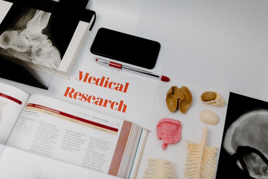
Synovial joints are the most mobile type of joint in the human body, enabling a wide range of movements such as flexion, extension, abduction, adduction, rotation, and circumduction. Their structure comprises a synovial cavity filled with lubricating fluid, articular cartilage covering bone ends, a fibrous capsule, synovial membrane, and various ligaments providing stability. The synovial fluid reduces friction, supplies nutrients, and removes waste, allowing smooth articulation. The capsule encloses the joint, maintaining the fluid and protecting the joint from external forces. Ligaments, both collateral and cruciate, anchor the joint and limit excessive motion, preventing dislocation. The articular cartilage, composed of type II collagen and proteoglycans, provides a low-friction, shock-absorbing surface that distributes load evenly across the joint. The synovial membrane secretes the fluid and repairs cartilage damage. Joint stability is achieved through a combination of passive structures (capsule, ligaments, cartilage) and active muscular control. Muscles contract to produce movement, while tendons transmit force from muscle to bone. The coordination of these elements allows precise, controlled motion essential for daily activities and athletic performance. Pathologies such as osteoarthritis, rheumatoid arthritis, and ligament sprains often arise from degeneration of cartilage, inflammation of the synovial membrane, or compromise of ligament integrity, leading to pain, reduced range of motion, and functional impairment. Understanding the mechanics of synovial joints is crucial for diagnosing joint disorders, designing rehabilitation protocols, and developing surgical interventions that restore joint congruity and function.
Future research targets biomimetic materials to repair joints, aiming to mimic resilience and low-friction surfaces, thus prolonging joint function.
Growth!!
Muscle Fiber Types
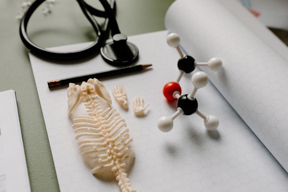
Muscle fibers are classified into Type I (slow‑oxidative), Type IIa (fast‑oxidative‑glycolytic), and Type IIb/x (fast‑glycolytic). Type I fibers excel in endurance, while Type II fibers generate rapid, powerful contractions. They are crucial for endurance and explosive power.!!
Skeletal Muscle Contraction
In skeletal muscle, contraction is initiated by a motor neuron releasing acetylcholine at the neuromuscular junction, depolarizing the sarcolemma and propagating an action potential along the T‑tubules. The depolarization triggers the ryanodine receptors on the sarcoplasmic reticulum to release Ca²⁺ into the cytosol. Calcium binds to troponin C, causing a conformational shift that moves tropomyosin away from actin’s myosin‑binding sites. Myosin heads, energized by ATP hydrolysis, form cross‑bridges with actin, executing the power stroke that shortens the sarcomere. The cycle repeats as long as Ca²⁺ remains elevated, allowing sustained contraction. Relaxation occurs when the sarcoplasmic reticulum actively pumps Ca²⁺ back, troponin‑tropomyosin re‑occludes the binding sites, and the muscle returns to its resting length. This sliding‑filament mechanism underlies all voluntary movements and is regulated by neural input, metabolic state, and hormonal signals. During tetanic stimulation, the frequency of action potentials exceeds the refractory period, producing a sustained rise in intracellular Ca²⁺ and a near‑maximal force output. The force generated is proportional to the number of motor units recruited and the firing rate, a principle known as the size principle. Additionally, the cross‑bridge cycle efficiency is modulated by phosphorylation of myosin light chains, which enhances ATPase activity and increases the rate of cross‑bridge detachment. The interplay between calcium dynamics, cross‑bridge cycling, and metabolic supply determines the contractile performance of skeletal muscle under various physiological demands. Furthermore, the elasticity of titin contributes to passive tension during stretch, allowing the muscle to return to its resting length without energy expenditure. The recruitment of motor units follows a hierarchical pattern, where smaller, fatigue‑resistant units are activated first, followed by larger, powerful units as force demands increase. Finally, the modulation of intracellular calcium by calcium‑binding proteins such as parvalbumin fine‑tunes contraction speed and fatigue resistance across different muscle fiber types. Collectively, these molecular and cellular mechanisms enable skeletal muscle to adapt to diverse mechanical loads, from fine motor tasks to maximal force production. The sarcoplasmic reticulum’s calcium‑handling proteins, such as SERCA pumps and calsequestrin, regulate the rate of Ca²⁺ reuptake, influencing relaxation speed and fatigue resistance. Moreover, the presence of myosin heavy‑chain isoforms determines the intrinsic velocity of cross‑bridge cycling, allowing specialized fibers to perform rapid contractions or sustain prolonged activity.
Connective Tissue Roles
Connective tissues—bone, cartilage, ligaments, tendons—provide structural support, protect organs, anchor muscles, and transmit forces. They also store minerals, mediate healing, and maintain joint stability through elastic and fibrous matrices and aid circulation.
Ligaments and Tendons
Ligaments are dense, fibrous connective tissues that connect bone to bone, providing joint stability and limiting excessive motion. Tendons attach muscle to bone, transmitting contractile forces to produce movement. Both structures are rich in collagen type I, giving them tensile strength and elasticity. Ligaments contain fibroblasts, fibrocytes, and a matrix of collagen fibers arranged in parallel bundles, allowing them to resist shear and tensile forces; Tendons have a hierarchical organization: collagen fibrils form fibers, fibers bundle into fascicles, and fascicles assemble into the tendon. This architecture enables efficient load transfer from muscle to bone. Ligaments also house mechanoreceptors such as Ruffini endings and Pacinian corpuscles, which contribute to proprioception and joint position sense. Tendons possess a high vascular supply through the paratenon, facilitating nutrient delivery and waste removal. Injury mechanisms differ: ligament sprains result from overstretching or tearing, while tendon ruptures often involve overuse or acute overload. Healing processes involve an inflammatory phase, a proliferative phase where fibroblasts synthesize new collagen, and a remodeling phase that reorganizes collagen fibers. Clinical management ranges from rest and immobilization to surgical reconstruction for severe tears. Understanding the biomechanical properties of ligaments and tendons is essential for diagnosing joint instability, planning orthopedic interventions, and designing rehabilitation protocols. During healing, the tendon’s collagen fibers realign along the line of force, improving tensile strength over months. Physical therapy often incorporates eccentric loading exercises to stimulate collagen remodeling and restore functional capacity. Rehabilitation focuses on restoring range of motion, strength, and proprioception!! for athletes!!!.
Common Disorders
Common disorders of the musculoskeletal system include osteoporosis, arthritis, muscular dystrophies, and tendonitis. These conditions affect bone density, joint cartilage, muscle fibers, and connective tissues, leading to pain, reduced mobility, and functional impairment. and increased of fractures.!!
Osteoporosis and Arthritis
Osteoporosis is a systemic skeletal disorder characterized by decreased bone mass and microarchitectural deterioration, increasing fracture risk. Risk factors include age, hormonal changes, low calcium intake, sedentary lifestyle, and certain medications. Diagnostic criteria rely on dual-energy X‑ray absorptiometry (DXA) T‑scores, with <‑2.5 indicating osteoporosis. Management focuses on calcium and vitamin D supplementation, weight‑bearing exercise, pharmacologic agents such as bisphosphonates, selective estrogen receptor modulators, and denosumab, alongside lifestyle modifications to reduce fall risk.
Arthritis encompasses a spectrum of joint diseases, primarily osteoarthritis (degenerative) and rheumatoid arthritis (autoimmune). Osteoarthritis features cartilage wear, subchondral bone sclerosis, osteophyte formation, and synovial inflammation, often presenting with pain, stiffness, and reduced joint function. Rheumatoid arthritis involves synovial hyperplasia, pannus formation, and systemic inflammation, leading to joint destruction and extra‑articular manifestations. Diagnosis employs clinical assessment, imaging (X‑ray, MRI), serologic markers (RF, anti‑CCP), and joint fluid analysis. Treatment strategies include NSAIDs, disease‑modifying antirheumatic drugs (DMARDs), biologics, physical therapy, and, in advanced cases, joint replacement surgery.

Patients often experience chronic pain and swelling, limiting mobility and requiring multidisciplinary caredaily.

Diagnostic Imaging Techniques
Diagnostic imaging assesses bone and soft tissue. X‑ray detects fractures and osteoarthritic changes, while MRI visualizes ligament tears, cartilage loss, and marrow edema. These modalities guide therapy, monitor progression, and help prevent complications. Early detection improves outcomes and reduces costs!
MRI and X-Ray Applications
Magnetic resonance imaging (MRI) offers unparalleled soft‑tissue contrast, enabling precise visualization of cartilage, ligaments, tendons, and intervertebral discs. It excels in diagnosing meniscal tears, rotator cuff ruptures, and occult fractures that radiographs may miss. MRI’s multiplanar capability allows clinicians to assess joint congruity, detect bone marrow edema, and evaluate the extent of osteonecrosis. In contrast, X‑ray imaging remains the first‑line modality for evaluating bone alignment, detecting fractures, and monitoring osteoarthritis progression. Radiographs provide rapid, cost‑effective assessment of cortical bone integrity and joint space narrowing, essential for initial trauma evaluation and long‑term follow‑up. When combined, MRI and X‑ray complement each other: X‑ray establishes bony anatomy, while MRI delineates soft‑tissue pathology. This dual‑modality approach informs surgical planning, guides arthroscopic interventions, and supports conservative management decisions. Emerging techniques such as low‑dose CT and advanced MRI sequences (e.g., dGEMRIC, T2 mapping) further refine diagnostic accuracy, allowing early detection of cartilage degeneration and subtle bone changes. Together, these imaging tools form a comprehensive diagnostic arsenal that enhances patient outcomes by facilitating timely, targeted treatment strategies. High‑resolution MRI sequences and quantitative bone density mapping give clinicians precise metrics to tailor rehabilitation and monitor treatment effectiveness over time ensuring optimal functional recovery daily